PUBLICATIONS
A full list of publications can be found on Google Scholar.
RESEARCH SPOTLIGHTS
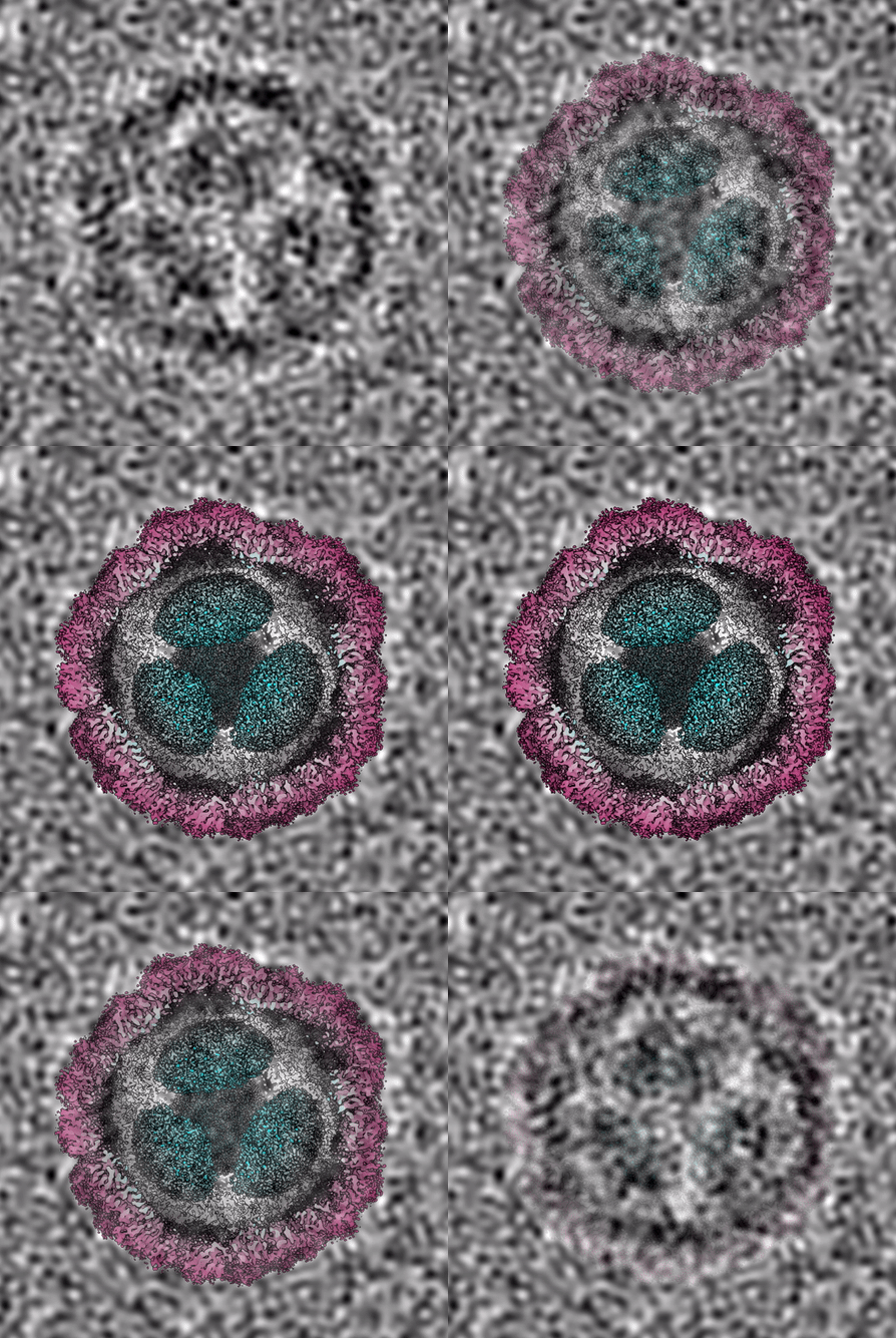
Pore dynamics and asymmetric cargo loading in an encapsulin nanocompartment
Science Advances 2022, 8(4), eabj4461.

Encapsulins are protein nanocompartments that house various cargo enzymes, including a family of decameric ferritin-like proteins. Here, we study a recombinant Haliangium ochraceum encapsulin:encapsulated ferritin complex using cryo–electron microscopy and hydrogen/deuterium exchange mass spectrometry to gain insight into the structural relationship between the encapsulin shell and its protein cargo. An asymmetric single-particle reconstruction reveals four encapsulated ferritin decamers in a tetrahedral arrangement within the encapsulin nanocompartment. This leads to a symmetry mismatch between the protein cargo and the icosahedral encapsulin shell. The encapsulated ferritin decamers are offset from the interior face of the encapsulin shell. Using hydrogen/deuterium exchange mass spectrometry, we observed the dynamic behavior of the major fivefold pore in the encapsulin shell and show the pore opening via the movement of the encapsulin A-domain.

Dr Jenn Ross

Dr Tom Lambert

Dr Kelly Gallagher
Conservation of the structural and functional architecture of encapsulated ferritins in bacteria and archaea. Biochem J. 2019, 476, 975-989. doi: 10.1042/BCJ20180922
Structural characterization of encapsulated ferritin provides insight into iron storage in bacterial nanocompartments. eLife 2016, 5, e18972. DOI: 10.7554/eLife.18972
Ferritins are ubiquitous proteins that oxidise and store iron within a protein shell to protect cells from oxidative damage. We have characterized the structure and function of a new member of the ferritin superfamily that is sequestered within an encapsulin capsid. We show that this encapsulated ferritin (EncFtn) has two main alpha helices, which assemble in a metal dependent manner to form a ferroxidase center at a dimer interface. EncFtn adopts an open decameric structure that is topologically distinct from other ferritins. While EncFtn acts as a ferroxidase, it cannot mineralize iron. Conversely, the encapsulin shell associates with iron, but is not enzymatically active, and we demonstrate that EncFtn must be housed within the encapsulin for iron storage. This encapsulin nanocompartment is widely distributed in bacteria and archaea and represents a distinct class of iron storage system, where the oxidation and mineralization of iron are distributed between two proteins.

RECENT AND NOTEWORTHY
81. Determining the Location of the Alpha-Synuclein Dimer Interface using Native Top-Down Fragmentation and Isotope Depletion-Mass Spectrometry. K. Jeacock, A. Chappard, K. Gallagher, C. Mackay, D. Kilgour, M. Horrocks, T. Kunath, D.J. Clarke. J. Am. Soc. Mass Spec. 2023. 34, 5, 847–856.
79. Single-Molecule Two-Color Coincidence Detection of Unlabeled alpha-Synuclein Aggregates
A. Chappard, C. Leighton, R.S. Saleeb, K. Jeacock, S.R. Ball, K. Morris, O. Kantelberg, J. Lee, E. Zacco, A. Pastore, M. Sunde, D.J. Clarke, P. Downey, T. Kunath, M.H. Horrocks. Angewandte Chemie International Edition, 2023, e202216771
78. A mechanistic evaluation of human beta defensin 2 mediated protection of human skin barrier in vitro
J. R. Shelley, B. J. McHugh, J. Wills, J. R. Dorin, R. Weller, D. J. Clarke, D. J. Davidson. Scientific Reports 2023, 13, 2271.
77. Design and Fabrication of a Fully-Integrated, Miniaturised Fluidic System for the Analysis of Enzyme Kinetics. A. Tsiamis, A. Buchoux, S. Mahon, A. Walton, S. Smith, D.J. Clarke, A. Stokes. Micromachines. 2023, 14 (3), 537.
76. Pathological structural conversion of α-synuclein at the mitochondria induces neuronal toxicity.
M Choi , A Chappard , B Singh , C Maclachlan , M Rodrigues , E Fedotova , A Berezhnov, S De, C Peddie, D Athauda, G Virdi, W Zhang, J Evans, A Wernick, Z Shadman Zanjani , P Angelova, N Esteras, A Vinokurov ,K Morris ,K Jeacock , L Tosatto , D Little , P Gissen , DJ Clarke, T Kunath, L Collinson, D Klenerman, A Abramov, M Horrocks, S Gandhi. Nature Neuroscience. 2022, 25 (9), 1134-1148.
75. Native mass spectrometry guided screening identifies hit fragments for HOP-HSP90 PPI inhibition. CGL Veale, M Vaaltyn, M Mateos-Jimenez, R Müller, CL Mackay, A Edkins, DJ Clarke. ChemBioChem. 2022, 23 (21), e202200322.
74. A ribosomally synthesized and post-translationally modified peptide containing a β-enamino acid and a macrocyclic motif, S. Wang, S. Lin, Q. Fang, R Gyampoh, Z Lu, Y Gao, DJ Clarke, K Wu, L Trembleau, Y Yu, K Kyeremeh, B Milne, J Tabudravu, H Deng. Nature Comms. 2022, 13(1), 5044.
73. Probing TDP-43 Condensation using an In Silico Designed Aptamer. E Zacco , O Kantelberg ,E Milanetti , A Armaos ,F Paolo Panei , J Gregory , K Jeacock , S Chandran , DJ Clarke , G Ruocco , S Gustincich , M Horrocks , A Pastore, GG Tartaglia. Nature Comms. 2022, 13(1), 3306.
72. Pore dynamics and asymmetric cargo loading in an encapsulin nanocompartment. J Ross, Z McIver, T Lambert, C Piergentili, J Bird, K J Gallagher, F L Cruickshank, P James , E Zarazúa-Arvizu, L E Horsfall, K J Waldron, M D Wilson, C L Mackay, A Baslé, D J Clarke*, J Marles-Wright*. Science Advances 2022, 8(4), eabj4461.
71. A native mass spectrometry platform identifies HOP inhibitors that modulate the HSP90-HOP protein-protein interaction. C G L Veale, M Mateos-Jimenez, M C Vaaltyn, R Müller, M P Makhubu, M Alhassan, B G de la Torre, F Albericio, C L Mackay, A L Edkins, D J Clarke. Chem Commun. 2021, 57, 10919-10922. doi: 10.1039/d1cc04257b.
70. Dissecting the structural and functional roles of a putative metal entry site in encapsulated ferritins
C. Piergentili, J. Ross, D. He, K. J. Gallagher, W. A. Stanley, L. Adam, C. L. Mackay, A. Baslé, K. J Waldron, D. J. Clarke*, J. Marles-Wright*. J. Biol. Chem., 2020, 295 (46), 15511-15526. https://doi.org/10.1074/jbc.RA120.014502
69. Isotope Depletion Mass Spectrometry (ID-MS) for Accurate Mass Determination and Improved Top-Down Sequence Coverage of Intact Proteins. K. J. Gallagher, M. Palasser, S. Hughes, C. L. Mackay, DPA Kilgour, D. J. Clarke. Journal of the American Society for Mass Spectrometry, 2020, 31 (3), 700-710. https://doi.org/10.1021/jasms.9b00119
68. Native Ion Mobility Mass Spectrometry (IM‐MS) reveals that small organic acid fragments impart gas‐phase stability to carbonic anhydrase II. C.G.L. Veale, M. Mateos Jimenez, C.L. Mackay, D.J. Clarke. Rapid Comms Mass Spec 2020, 34 (2), e8570. https://doi.org/10.1002/rcm.8570.
67. High resolution fourier transform ion cyclotron resonance mass spectrometry (FT-ICR MS) for the characterisation of enzymatic processing of commercial lignin. V. Echavarri-Bravo, M. Tinzl, W. Kew, F. Cruickshank, C. L. Mackay, D. J. Clarke, L. E. Horsfall. New biotechnology. 2019, 52, 1-8. doi.org/10.1016/j.nbt.2019.03.001
66. Untargeted Metabolite Mapping in 3D Cell Culture Models Using High Spectral Resolution FT-ICR Mass Spectrometry Imaging. L. H. Tucker, G. R. Hamm, R. J. E. Sargeant, R. J.A. Goodwin, C. L. Mackay, C. J. Campbell, D. J. Clarke. Analytical Chem. 2019, 91, 9522-9529. doi.org/10.1021/acs.analchem.9b00661
65. Conservation of the structural and functional architecture of encapsulated ferritins in bacteria and archaea.
D. He, C. Piergentili, J. Ross, E. Tarrant, L. R. Tuck, C. L. Mackay, Z. McIver, K. J. Waldron, J. Marles-Wright*, D. J. Clarke*. Biochem J. 2019, 476, 975-989. doi: 10.1042/BCJ20180922
64. Use of isotopically labeled substrates reveals kinetic differences between human and bacterial serine palmitoyltransferase. P. J. Harrison, K. Gable, N. Somashekarappa, V. Kelly, D. J. Clarke, J. H. Naismith, T. M. Dunn, D. J. Campopiano. J. Lipid Research. 2019, 60, 953-962. doi: 10.1194/jlr.M089367.
63. S-nitrosylation of the zinc finger protein SRG1 regulates plant immunity. B. Cui, Q. Pan, D. J. Clarke, M. Ochoa Villarreal, S. Umbreen, B. Yuan, W. Shan, J. Jiang & G. J. Loake. Nature Comms. 2018, 9(1), 4226. doi: 10.1038/s41467-018-06578-3.
W. Kew, C. L. Mackay, I. Goodall, D. J. Clarke*, and D. Uhrin*. Anal. Chem. 2018, 90, 11265-11272. doi: 10.1021/acs.analchem.8b01446.
61. MALDI Matrix Application Utilizing a Modified 3D Printer for Accessible High Resolution Mass Spectrometry Imaging. L. Tucker, A. Conde-González, D. Cobice, G. Hamm, R. J. A. Goodwin, C. J. Campbell, D. J. Clarke, C. L. Mackay. Anal. Chem. 2018, 90, 8742–8749. doi: 10.1021/acs.analchem.8b00670
60. Autopiquer - a Robust and Reliable Peak Detection Algorithm for Mass Spectrometry. D. P. A. Kilgour, S. Hughes, S. L. Kilgour, C. L. Mackay, M. Palmblad, B. Q. Tran, Y. Ah Goo, R. K. Ernst, D. J. Clarke, D. R. Goodlett. J. Am. Soc. Mass Spec. 2017, 28, 253-262. doi: 10.1007/s13361-016-1549-z.
59. Chemical Diversity and Complexity of Scotch Whisky as Revealed by High-Resolution Mass Spectrometry. W. Kew, I. Goodall, D. J. Clarke*, D. Uhrin*. J. Am. Soc. Mass Spec. 2017, 28, 200-213. doi:10.1007/s13361-016-1513-y.
58. Interactive van Krevelen diagrams – Advanced visualisation of mass spectrometry data of complex mixtures. W. Kew, J. Blackburn, D. J. Clarke, D. Uhrin. Rapid Comms. Mass. Spec. Volume 31, 2017, 7, 658–662. doi: 10.1002/rcm.7823.
57. L-1 beta-induced protection of keratinocytes against Staphylococcus aureus-secreted proteases is mediated by human beta defensin 2. B. Wang, B. J. McHugh, A. Qureshi, D. J. Campopiano, D.J. Clarke, J. R. Fitzgerald,J. Dorin, R. Weller, D. J. Davidson. J Invest. Dermatol. 2017,137, 95–105. doi: 10.1016/j.jid.2016.08.025.
56. Characterisation of homologous sphingosine 1-phosphate lyase (S1PL) isoforms in the bacterial pathogen Burkholderia pseudomallei. C. McLean, J. Marles-Wright, R. Custodio,J. Lowther, A. J. Kennedy, J. Pollock, D. J. Clarke, A. R. Brown, D. J. Campopiano. J. Lipid Res. 2017, 58, 137-150. doi: 10.1194/jlr.M071258.
55. Structural characterization of encapsulated ferritin provides insight into iron storage in bacterial nanocompartments. D. He, S. Hughes, S. Vanden-Hehir, A. Georgiev, K. Altenbach, E. Tarrant, C. L. Mackay, K. J. Waldron, D. J. Clarke*, J. Marles-Wright*. eLife 2016, 5:e18972. doi: 10.7554/eLife.18972
54. Determination of protein disulfide bond reduction potential by isotope labelling and intact mass measurement. S. E. Thurlow, D. P. Kilgour, D. J. Campopiano, C. L. Mackay, P. R. R. Langridge-Smith, D. J. Clarke*, C. J. Campbell*. Anal Chem 2016, 88, 2727–2733. doi: 10.1021/acs.analchem.5b04195.
53. The crystal structure of the propionaldehyde dehydrogenase enzyme from Clostridium phytofermentans with NAD+ and CoA in the active site provides insight into cofactor binding and the mechanism of acyl-transfer in acylating aldehyde dehydrogenase enzymes. L. R. Tuck, K. Altenbach, A. T. Fu, A. D. Crawshaw, D. J. Campopiano, D. J. Clarke, and J. Marles-Wright. Scientific Reports. 2016, 6, 22108. doi:10.1038/srep22108.
52. Mass spectrometry analysis of the oxidation states of the pro-oncogenic protein anterior gradient-2 reveals covalent dimerization via an intermolecular disulphide bond. D. J. Clarke*, E. Murray, J. Faktor, A. Mohtar, B. Vojtesek, C. L. MacKay, P. Langridge Smith, T. Hupp*. BBA: Proteins and Proteomics 2016, 1864 551-561.
doi:10.1016/j.bbapap.2016.02.011.
51. New cytotoxic callipeltins from the Solomon Island marine sponge Asteropus sp. M. Stierhof, K. Ø. Hansen, M. Sharma, K. Feussner, K. Subko, F. F. Díaz-Rullo, J. Isaksson, I. Pérez-Victoria, D. J. Clarke, E. Hansen, M Jaspars, J. N. Tabudravua. Tetrahedron 2016, 72 (44), 6929-6934. Link.
50. Characterization of secreted sphingosine-1-phosphate lyases required for virulence and intracellular survival of Burkholderia pseudomallei. R. Custódio, C. J. McLean, A. E. Scott, J. Lowther, A. Kennedy, D. J. Clarke, D. J. Campopiano, M. Sarkar-Tyson, A. R. Brown. Mol Microbiol. 2016, 102, 1004-1019. doi: 10.1111/mmi.13531
49. Molecular basis of Streptococcus mutans sortase A inhibition by the flavonoid natural product trans-chalcone. D. J. Wallock-Richards, J. Marles-Wright, D. J. Clarke, A. Maitra, M. Dodds, B. Hanley, D. J. Campopiano. Chem Comm. 2015, 51, 10483-10485. doi: 10.1039/C5CC01816A.
48. Desalting Large Protein Complexes during Native Electrospray Mass Spectrometry by Addition of Amino Acids to the Working Solution. D. J. Clarke*, D. J. Campopiano. Analyst, 2015, 140, 2679-2686. doi: 10.1039/C4AN02334J
47. Insights into the Conformations of Three Structurally Diverse Proteins: Cytochrome c, p53, and MDM2, Provided by Variable-Temperature Ion Mobility Mass Spectrometry. E. R. Dickinson, E. Jurneczko, K. J. Pacholarz, D. J. Clarke, M. Reeves, K. L. Ball, T. Hupp, D. J. Campopiano, P. V. Nikolova, and P. E. Barran. Anal Chem 2015, 87, 3231-3238.
46. Dissecting the dynamic conformations of the metamorphic protein lymphotactin. Harvey, S.R., Porrini, M., Konijnenberg, A., Clarke, D.J., Tyler, R.C., Langridge-Smith, P.R., MacPhee, C.E., Volkman, B.F., Barran, P.E. J. Phy. Chem. B, 2014, 118, 12348-12359.
45. The chemical basis of serine palmitoyltransferase inhibition by myriocin. D. J. Clarke, J. M. Wadsworth, S. A. McMahon, J. P. Lowther, A. E. Beattie, H. Broughton, T. M. Dunn, J. H. Naismith, and D. J. Campopiano. J. Am. Chem. Soc., 2013, 135, 14276-14285. DOI: 10.1021/ja4059876
44. Probing the conformational diversity of cancer-associated mutations in p53 by Ion Mobility-Mass Spectrometry. E. Jurneczko, F. Cruickshank, M. Porrini, D. J. Clarke, I. D. G. Campuzano, M. Morris, P. V. Nikolova, and P. E. Barran. Angewandte Chemie Int. Ed., 2013, 52, 4370-4374.
43. Redox Regulation of Tumour Suppressor Protein p53: Identification of the Sites of Hydrogen Peroxide Oxidation and Glutathionylation. D. J. Clarke, J. Scotcher, C. L. Mackay, T. Hupp, P. J. Sadler, and P. R. R. Langridge-Smith. Chemical Science, 2013, 4, 1257-1269. DOI: 10.1039/C2SC21702C
42. Reconstitution of the pyridoxal 5’-phosphate (PLP) dependent enzyme serine palmitoyltransferase (SPT) with pyridoxal reveals a crucial role for the phosphate during catalysis. A. E. Beattie, D. J. Clarke, J. M. Wadsworth, J. Lowther, H. Sin and D. J. Campopiano. Chem. Comm., 2013, 49, 7058.
31. Mapping a Non-covalent Protein-Peptide Interface by Top-Down FT-ICR Mass Spectrometry using Electron Capture Dissociation. D. J. Clarke, E. Murray, T. Hupp, C. L. Mackay, and P. R. R. Langridge-Smith. J. Am. Soc. Mass Spec. 2011, 22 ,1432-1440.
30. Identification of Two Reactive Cysteine Residues in the Tumor Suppressor Protein p53 Using Top-Down FTICR Mass Spectrometry. J. Scotcher, D. J. Clarke, S. K. Weidt, C. L. Mackay, T. R. Hupp, P. J. Sadler, P. R. R. Langridge-Smith. J. Am. Soc. Mass Spec. 2011, 22, 888-897.
29. Online Quench-Flow Electrospray Ionization Fourier Transform Ion Cyclotron Resonance Mass Spectrometry for Elucidating Kinetic and Chemical Enzymatic Reaction Mechanisms. D. J. Clarke, A. A. Stokes, P. R. R. Langridge-Smith, C. L. Mackay. Anal. Chem. 2010, 82, 1897-1904.
28. Subdivision of the bacterioferritin comigratory protein family of bacterial peroxiredoxins based on catalytic activity. D. J. Clarke, X. P. Ortega, C. L. Mackay, M. A. Valvano, J. Govan, D. J. Campopiano, P. Langridge-Smith and A. R. Brown. Biochemistry. 2010, 49, 1319-1330.
Redox Regulation of Tumour Suppressor Protein p53: Identification of the Sites of Hydrogen Peroxide Oxidation and Glutathionylation. Chemical Science, 2013, 4, 1257-1269. DOI: 10.1039/C2SC21702C

The p53 transcription factor is a key tumour suppressor protein. In this paper we analyze oxidation pathways in the p53 core domain by high resolution mass spectrometry and top-down fragmentation. Firstly, we show that p53 core domain is sensitive to oxidation by the reactive oxygen species (ROS) and that the zinc-coordination site is the initial target for ROS-induced oxidation. Two disulfide bonds are formed involving Cys182 and the three cysteines which coordinate to zinc (Cys176, 238 and 242). This disulfide bond formation is accompanied by loss of zinc from the binding site. Our work also highlights an additional cysteine, Cys277, is prone to oxidation via a ROS-independent mechanism. We discuss our findings in the context of redox regulation of p53 activity and in comparison to other redox regulated proteins.
